Date:16/02/16
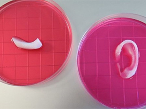 Scientists have developed an innovative 3D bioprinter capable of generating replacement tissue that’s strong enough to withstand transplantation. To show its power, the scientists printed a jaw bone, muscle, and cartilage structures, as well as a stunningly accurate human ear.
After nearly 10 years in development, a research team led by Anthony Atala from Wake Forest Institute for Regenerative Medicine has unveiled the Integrated Tissue and Organ Printing System (ITOP). Once refined and proven safe in humans, these 3D bioprinted structures could be used to replace injured, missing, or diseased tissue in patients. And because they’re designed in a computer, these replacement parts will be made to order to meet the unique needs of each patient. The details of this breakthrough were published today in Nature Biotechnology.
Bioprinters work the same way that conventional 3D printers do, using additive manufacturing to build complex structures layer by layer. But instead of using plastics, resins, and metals, bioprinters use special biomaterials that closely approximate functional, living tissue.
But existing bioprinters cannot fabricate tissues of the right size or strength. Their products end up being far too weak and structurally unstable for surgical transplantation. They also cannot print more delicate structures like blood vessels, or vasculature. Without these ready-made blood vessels, cells cannot be supplied with critical nutrients and oxygen.
“Cells simply cannot survive without a blood vessel supply that’s smaller than 200 microns [0.07 inches], which is extremely small,” Atala told Gizmodo. “That’s the maximum distance. And that’s not just for printing, that’s nature.” He said it’s the “limiting factor” that has made bioprinting a particularly challenging technological proposition.
The new bioprinting system overcomes each of these shortcomings. Biodegradable plastic-like (polymer) materials are used to form the tissue shape, and a water-based gel delivers the cells to the structure (the gels aren’t toxic to the cells). A temporary outer structure helps to maintain the object’s shape during the printing process. To address the size limit, the researchers embedded microchannels into the design that allow nutrients and oxygen to be transported to cells anywhere within the structure.
“We basically recreated capillaries, creating microchannels that acted like a capillary bed,” said Atala.
Atala said his team’s 3D-printed tissues appear to have the right size, strength, and function for use in humans. Their system can generate human-scale, structurally stable tissues in virtually any shape, and parts can be modeled in a computer according to the precise physical needs of a patient.


New 3D bio-printer can make full size ear, muscle, and bone tissues
Scientists have developed an innovative 3D bioprinter capable of generating replacement tissue that’s strong enough to withstand transplantation. To show its power, the scientists printed a jaw bone, muscle, and cartilage structures, as well as a stunningly accurate human ear.After nearly 10 years in development, a research team led by Anthony Atala from Wake Forest Institute for Regenerative Medicine has unveiled the Integrated Tissue and Organ Printing System (ITOP). Once refined and proven safe in humans, these 3D bioprinted structures could be used to replace injured, missing, or diseased tissue in patients. And because they’re designed in a computer, these replacement parts will be made to order to meet the unique needs of each patient. The details of this breakthrough were published today in Nature Biotechnology.
Bioprinters work the same way that conventional 3D printers do, using additive manufacturing to build complex structures layer by layer. But instead of using plastics, resins, and metals, bioprinters use special biomaterials that closely approximate functional, living tissue.
But existing bioprinters cannot fabricate tissues of the right size or strength. Their products end up being far too weak and structurally unstable for surgical transplantation. They also cannot print more delicate structures like blood vessels, or vasculature. Without these ready-made blood vessels, cells cannot be supplied with critical nutrients and oxygen.
“Cells simply cannot survive without a blood vessel supply that’s smaller than 200 microns [0.07 inches], which is extremely small,” Atala told Gizmodo. “That’s the maximum distance. And that’s not just for printing, that’s nature.” He said it’s the “limiting factor” that has made bioprinting a particularly challenging technological proposition.
The new bioprinting system overcomes each of these shortcomings. Biodegradable plastic-like (polymer) materials are used to form the tissue shape, and a water-based gel delivers the cells to the structure (the gels aren’t toxic to the cells). A temporary outer structure helps to maintain the object’s shape during the printing process. To address the size limit, the researchers embedded microchannels into the design that allow nutrients and oxygen to be transported to cells anywhere within the structure.
“We basically recreated capillaries, creating microchannels that acted like a capillary bed,” said Atala.
Atala said his team’s 3D-printed tissues appear to have the right size, strength, and function for use in humans. Their system can generate human-scale, structurally stable tissues in virtually any shape, and parts can be modeled in a computer according to the precise physical needs of a patient.
Views: 547
©ictnews.az. All rights reserved.
Similar news
- The mobile sector continues its lead
- Facebook counted 600 million active users
- Cell phone testing laboratory is planned to be built in Azerbaijan
- Tablets and riders outfitted quickly with 3G/4G modems
- The number of digital TV channels will double to 24 units
- Tax proposal in China gets massive online feedback
- Malaysia to implement biometric system at all entry points
- Korea to build Green Technology Centre
- Cisco Poised to Help China Keep an Eye on Its Citizens
- 3G speed in Azerbaijan is higher than in UK
- Government of Canada Announces Investment in Green Innovation for Canada
- Electric cars in Azerbaijan
- Dominican Republic Govt Issues Cashless Benefits
- Spain raises €1.65bn from spectrum auction
- Camden Council boosts mobile security